
Click the thumbnails above to see images and captions
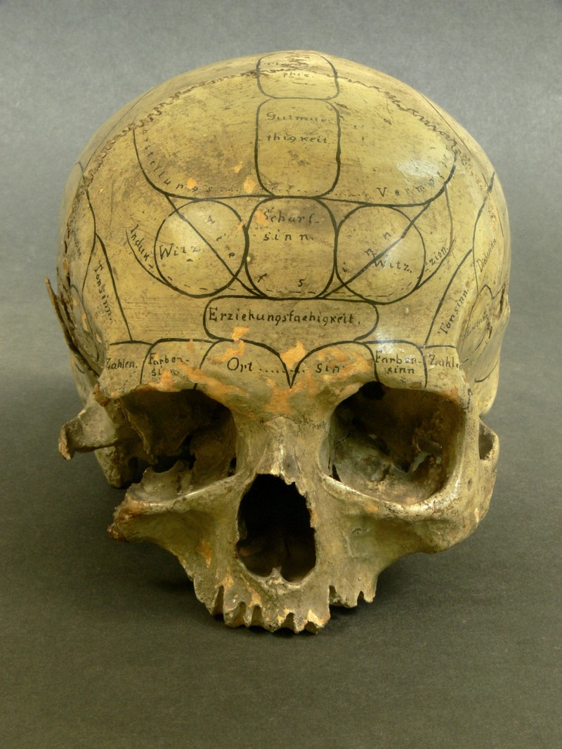
HUMAN SKULL INSCRIBED BY A PHRENOLOGIST Anonymous, nineteenth century. Photograph by Eszter Blahak/Semmelweis Museum.
Historically, the primary concern of neuroscience has been location. In the mass of flesh that is the human body, where is the mind? Towards the end of the eighteenth century, Franz Joseph Gall built an influential theory that posited that distinct areas of the cerebrum serve distinct faculties such as emotions, moral impulses and the intellect. He also thought that these different brain areas grow and shrink according to their use, pushing out and creating bumps on the skull that betray the makeup an individual's mind. It is easy today to chalk up Gall's reasoning to quackery, and its service to subsequent commercial and ideological uses (such as racism) certainly does not help his case. But Gall ushered in the first modern theory ascribing different mental functions to different parts of the cerebrum. While he was entirely off the mark on the details, his paradigm guides us to this day -- only the coordinate system has shifted to indicate positions within the brain rather than on the skull.
Portraits of the Mind: Visualizing the Brain from Antiquity to the 21st Century, by Carl Schoonover, foreword by Jonah Lehrer, is published by Abrams.
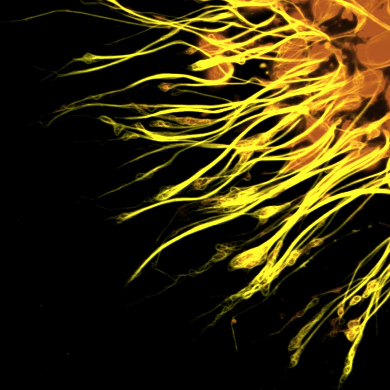
ANTIBODY STAIN Scaffolding in axons. Michael Hendricks and Suresh Jesuthasan (2008).
Antibody staining techniques take advantage of the exquisitely sensitive and selective properties of antibodies, the henchmen of the immune response. They can recognize, and strongly latch on to, molecules introduced from outside an organism's body, such as those lining the surface of pathogens. Molecular biologists have pioneered ways of harnessing the powerful ability of antibodies to recognize specific molecules and can employ them to study any protein of interest in the brain. By revealing where a given protein is found in a tissue and even within an individual cell, scientists are afforded precious insight into a rich molecular world otherwise invisible even under the microscope. This photomicrograph was generated using antibody that stains the molecular scaffolding found inside of axons, here in neurons that were growing in a dish.
Portraits of the Mind: Visualizing the Brain from Antiquity to the 21st Century, by Carl Schoonover, foreword by Jonah Lehrer, is published by Abrams.
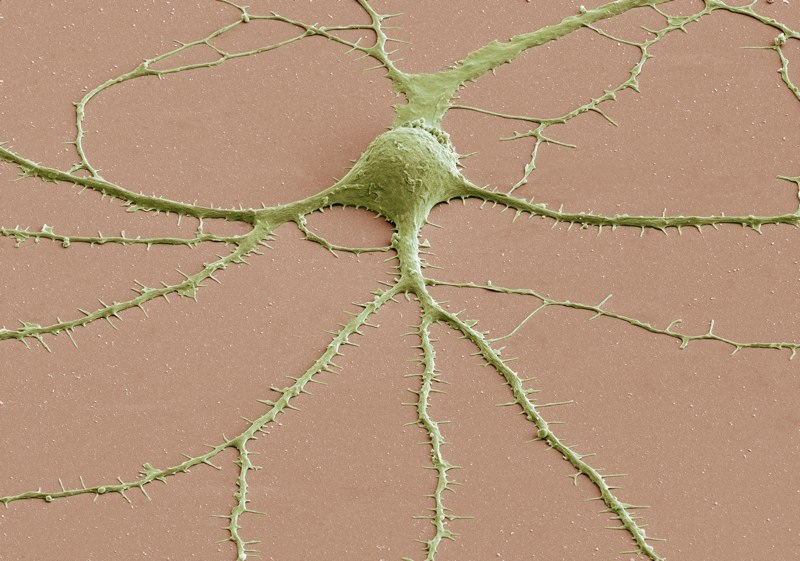
SCANNING ELECTRON MICROSCOPY Spiny neuron. Thomas Deerinck and Mark Ellisman (2009).
Electron microcopy grants researchers and clinicians access to a universe that is too small to be detected using light-based microscopes. This photomicrograph was obtained by scanning a beam of electrons across the sample while a detector kept track of electrons bouncing off its surface, betraying the specimen's outer shape. It reveals a neuron with its round-ish soma at the center, and thin dendrites radiating out of it. Pseudocoloring helps to differentiate elements in the image: The brown background is a surface of supportive glial cells, upon which the beige-colored neurons were grown before imaging.
Portraits of the Mind: Visualizing the Brain from Antiquity to the 21st Century, by Carl Schoonover, foreword by Jonah Lehrer, is published by Abrams.
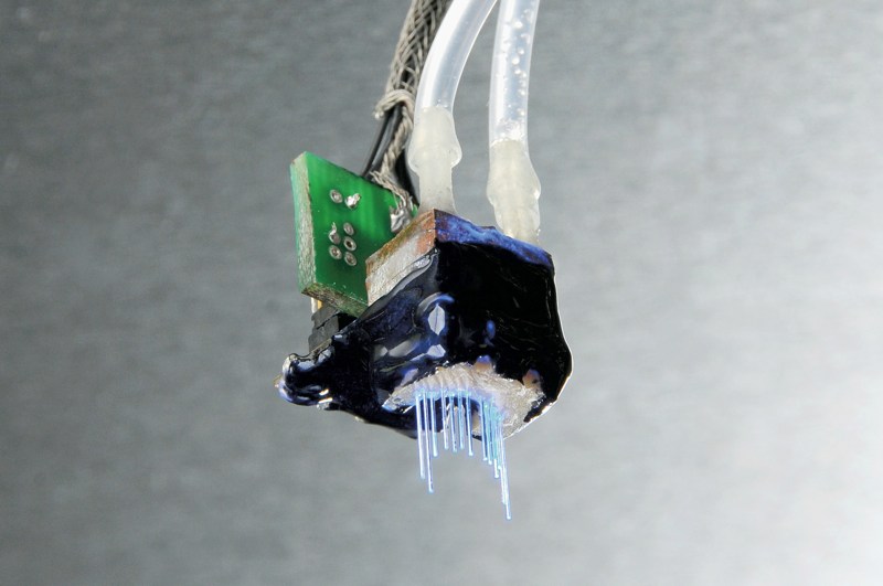
OPTICAL FIBER ARRAY Jacob Bernstein, Alexander Guerra, and Ed Boyden (2010).
A family of newly characterized proteins has been taking the neuroscience world by storm lately, due to their ability to turn light into electrical currents. Thanks to this property, researchers can switch neurons either on or off at will, using nothing but a fiber optic and a powerful light bulb. Early experiments that delivered these proteins to specific classes of neurons have restored vision to blind mice, and mitigated symptoms of Parkinson's disease in a rodent model of the disease; the human therapeutic applications, still far off in the future, are no less tantalizing.
With the imaginations of neuroscientists the world over running wild, the field is faced with serious engineering challenges--most critically, how to target beams of light to the right neurons. Engineers have been hard at work to design devices such as this one--a miniature array of independently controllable LED-coupled optical fibers. It is light enough to rest on top of a mouse or rat's head, and enables researchers to deliver light through its thin optical fibers to specific subsets of neurons in the animal's hippocampus. The goal is to causally investigate this brain area's mechanism with ever-increasing precision by selectively switching different parts of it on or off.
Portraits of the Mind: Visualizing the Brain from Antiquity to the 21st Century, by Carl Schoonover, foreword by Jonah Lehrer, is published by Abrams.
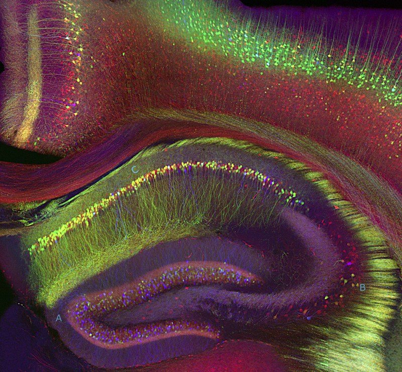
HIPPOCAMPUS Broad overview. Tamily Weissman, Jeff Lichtman, and Joshua Sanes (2005).
The late neurological patient known as "H. M." provided invaluable insight into human cognition when his debilitating epilepsy was treated half a century ago by the surgical removal of large portions of his hippocampus. While cured of his seizures, he also lost his ability to form long-term memories. Astonishingly, he could acquire and deploy new motor skills yet could not recall having learned them. His plight yielded compelling evidence for the view that different forms of memory are handled by different areas of the brain--that specific regions, such as the hippocampus, serve specific functions like conscious memory.
Yet region is itself a universe unto its own, and performs its assigned function thanks to a massively complex sub-circuit. This photomicrograph of a mouse hippocampus (bottom half) was fluorescently labeled so that different classes of neurons were illuminated in a specific color. (The colored labels are actually natural proteins that fluorescence on their own; researchers have simply introduced their genes into the neurons and let Nature do the rest of the work.)
Portraits of the Mind: Visualizing the Brain from Antiquity to the 21st Century, by Carl Schoonover, foreword by Jonah Lehrer, is published by Abrams.
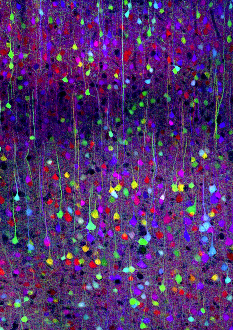
Neocortex Tamily Weissman, Jeff Lichtman, and Joshua Sanes (2007).
This photomicrograph of the mouse neocortex was obtained in transgenic mouse called "Brainbow", which employs a few different genetically-encoded fluorescent labels and mixes them up to create compound colors from a set of primaries. The mouse, which owes its existence to a breathtakingly elegant molecular biology magic trick (explained in detail in the book), allows researchers to light up cells in up to a hundred separate hues. This allows neuroscientists to distinguish adjacent neurons from one another and tease apart how they are connected to each other.
Portraits of the Mind: Visualizing the Brain from Antiquity to the 21st Century, by Carl Schoonover, foreword by Jonah Lehrer, is published by Abrams.
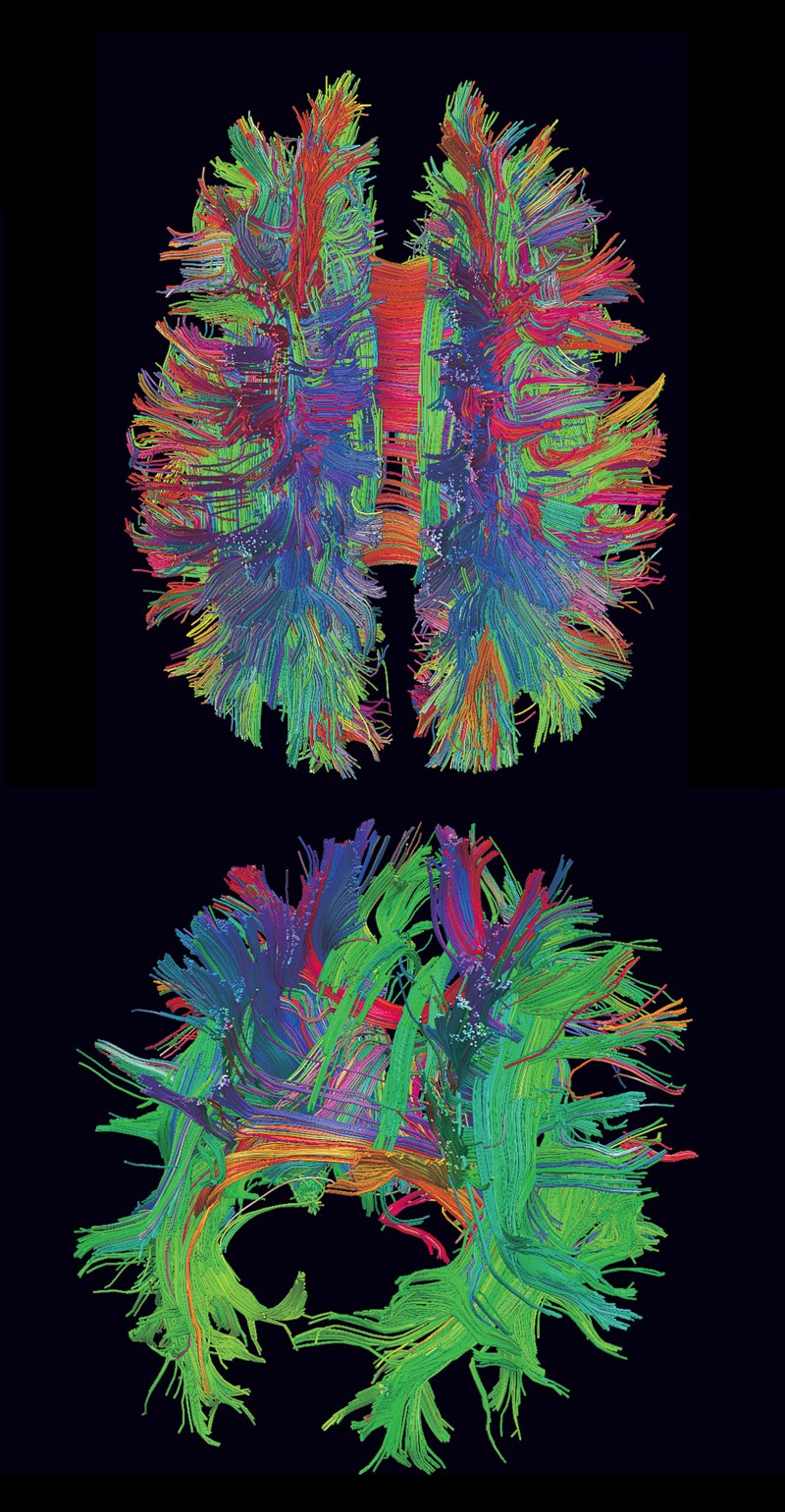
Diffusion MRI Patric Hagmann (2006).
A new method called diffusion MRI can be used to uncover major axon pathways in the brain by measuring the motion of water molecules contained within a group (or tract) of axons traveling from one point of the organ to another. This technique is capable of detecting water's natural diffusion along these tracts and thereby infers their paths indirectly.
This 3D reconstruction from diffusion MRI data shows a tractography of a human brain obtained in a live human subject who walked out of the apparatus unharmed. At top, we are looking down on the brain, with the back of the head at the bottom of the image and the forehead at top; in the view below it, we are looking at the subject from the back of the head. Each line does not represent a single axon, but thousands of them, traveling together as a group. The colors indicate the axis of each fiber (green: front to back; red: left to right; blue: top to bottom).
Portraits of the Mind: Visualizing the Brain from Antiquity to the 21st Century, by Carl Schoonover, foreword by Jonah Lehrer, is published by Abrams.
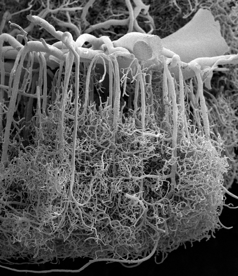
MICROVASCULATURE Opposite: Human cerebral cortex. Alfonso Rodríguez-Baeza and Marisa Ortega-Sánchez (2009). This photomicrograph shows a mouse brain whose blood vessels have been injected with India ink to fill and stain them. The brain was then automatically sliced and simultaneously imaged using a custom-designed microscope. This image, in which the blood vessels are represented in white for clarity, is a view from the front of a brain that has been sectioned through the middle.
Portraits of the Mind: Visualizing the Brain from Antiquity to the 21st Century, by Carl Schoonover, foreword by Jonah Lehrer, is published by Abrams.
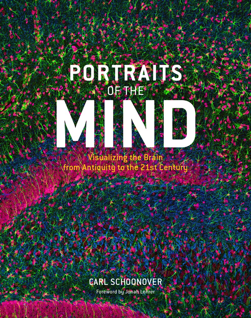
Portraits of the Mind follows the fascinating history of our exploration of the brain through images, from medieval sketches and 19th-century drawings by the founder of modern neuroscience to images produced using state-of-the-art techniques, allowing us to see the fantastic networks in the brain as never before. These black-and-white and vibrantly colored images, many resembling abstract art, are employed daily by scientists around the world, but most have never before been seen by the general public. Each chapter addresses a different set of techniques for studying the brain as revealed through the images, and each is introduced by a leading scientist in that field of study. Author Carl Schoonover's captions provide detailed explanations of each image as well as the major insights gained by scientists over the course of the past 20 years. Accessible to a wide audience, this book reveals the elegant methods applied to study the mind, giving readers a peek at its innermost workings, helping us to understand them, and offering clues about what may lie ahead.
Portraits of the Mind: Visualizing the Brain from Antiquity to the 21st Century, by Carl Schoonover, foreword by Jonah Lehrer, is published by Abrams.


No comments:
Post a Comment